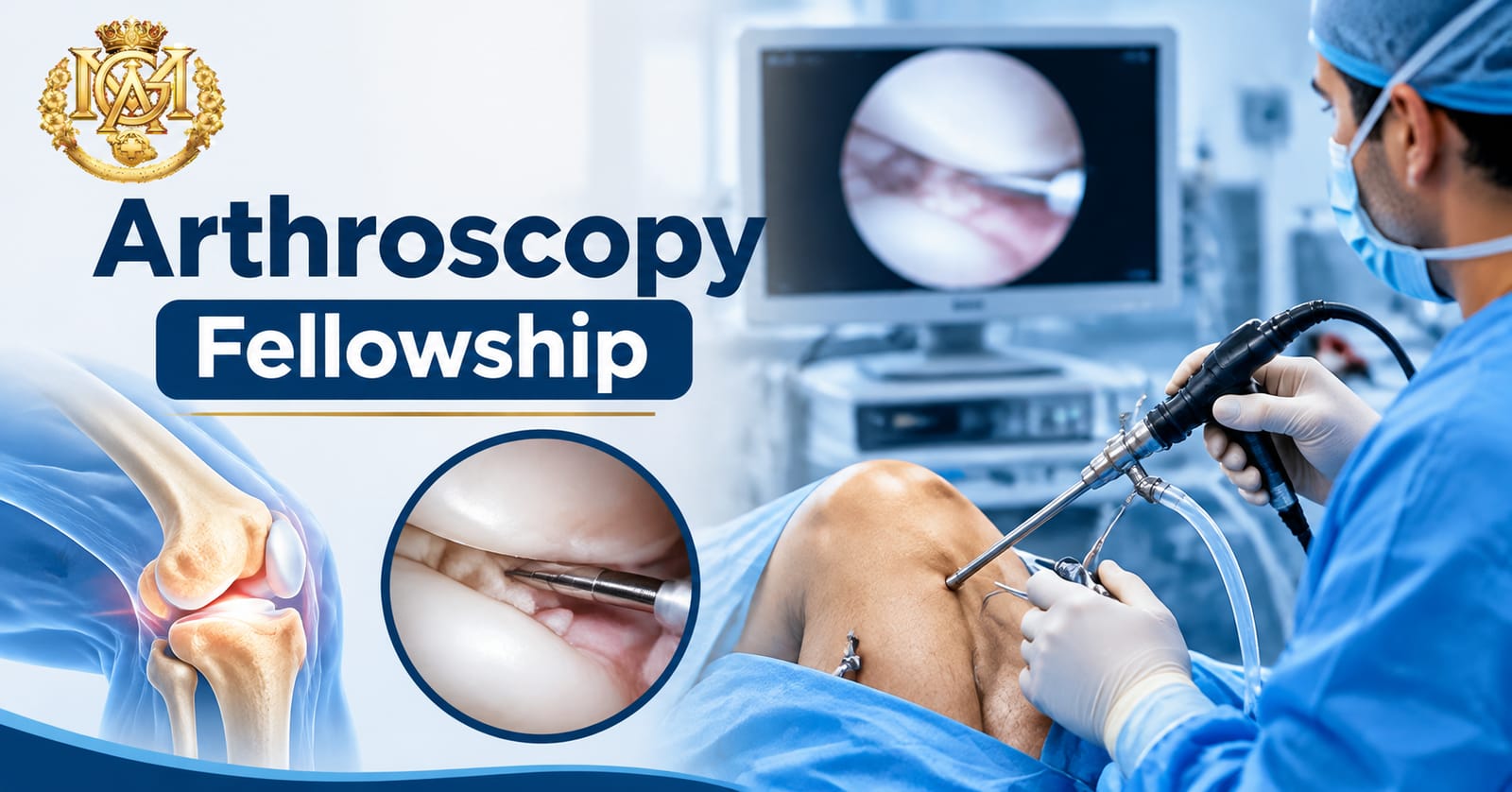
Arthroscopy is built on a detailed understanding of joint anatomy, biomechanics, and pathology. The instrument technique — the portal placement, the camera navigation, the operative steps — matters. But what makes an arthroscopic surgeon effective rather than merely technically proficient is the clinical science behind what they are seeing and doing inside the joint. Arthroscopy fellowship training systematically builds this scientific foundation alongside the operative skills. This article covers the core joint science that fellows study — the anatomy, the biomechanics, and the pathology — and why it matters for clinical practice. Doctors considering structured fellowship training can review the Fellowship in Arthroscopy and Sports Medicine at Medical Global Academy.
Why Joint Anatomy Is the Foundation of Arthroscopic Practice
Arthroscopy is a minimally invasive technique that accesses a joint through small portals and navigates it with a camera. This means that the surgeon's view is a close, restricted, and often distorted image of the internal joint environment. Making sense of that image — identifying normal from abnormal, mapping the anatomy the instrument is passing through, correlating what is seen with what was shown on pre-operative imaging — requires a precise and three-dimensional understanding of joint anatomy that goes beyond what standard medical education provides.
Fellowship training in arthroscopy and sports medicine spends significant time on joint anatomy for this reason. The anatomy is not reviewed as background material — it is studied as the operational map for everything the fellow will do in the operating theatre. Portal placement, instrument angles, and operative sequence are all derived from anatomical knowledge.
The Knee: The Highest-Volume Joint
Arthroscopy knee surgery accounts for the largest share of arthroscopic procedures globally. The knee is a complex synovial joint with multiple compartments, and fellowship training covers each of them in detail.
The medial and lateral compartments contain the femoral condyles, tibial plateaus, and the menisci. The medial and lateral menisci are fibrocartilaginous structures that distribute load across the knee, deepen the articular surface, and contribute to joint stability. Meniscal anatomy — the vascular zones, the attachment points, the difference between a repairable tear and one that requires resection — is a core component of fellowship study. The clinical implications are direct: a vascular peripheral tear in a young patient is a repair candidate; an avascular inner-zone tear in an older patient with degenerative changes is managed differently.
The intercondylar notch contains the anterior and posterior cruciate ligaments. ACL anatomy — the two bundles, the femoral and tibial footprints, the relationship of the ligament to the notch walls — is studied in depth because accurate graft placement in ACL reconstruction depends on reproducing the anatomical footprint. Fellowship training covers the evidence on anatomical versus non-anatomical tunnel placement and what it means for rotational stability post-reconstruction. For a clinical overview of ACL injury and its management, this article on ACL tear treatment covers the key considerations.
The patellofemoral compartment covers the relationship between the patella and the femoral trochlea. Patellofemoral pathology — chondral damage, patellar instability, trochlear dysplasia — is encountered regularly in sports medicine practice, and its management requires understanding the biomechanics of the extensor mechanism and the alignment forces that act on the patellofemoral joint.
Cartilage: The Science of Joint Preservation
Articular cartilage is avascular, aneural, and has limited capacity for self-repair. These properties make cartilage pathology one of the central challenges in sports medicine and one of the core science areas in fellowship training in musculoskeletal medicine.
Fellows study the structure of articular cartilage — the zonal organization of chondrocytes and collagen fibers, the proteoglycan matrix, and the relationship between cartilage structure and mechanical function. This is not abstract basic science: it directly informs clinical decision-making about which cartilage lesions are amenable to restoration procedures and which are not.
Cartilage lesion grading (Outerbridge, ICRS) is a standard tool in arthroscopic assessment. A Grade I lesion — cartilage softening — is managed differently from a Grade IV lesion with exposed subchondral bone. Fellowship training ensures that fellows can grade lesions consistently and reason through the management options: microfracture, osteochondral autograft transfer (OATS), autologous chondrocyte implantation (ACI), or conservative management depending on lesion size, location, patient age, and activity level.
Meniscal cartilage is a separate but related topic. The medial and lateral menisci are not articular cartilage, but their integrity is directly linked to articular cartilage health. Loss of meniscal tissue — through injury or resection — accelerates articular cartilage degeneration in the affected compartment. Fellowship training on meniscal repair versus meniscectomy is built on this understanding.
The Shoulder: Different Anatomy, Different Challenges
The shoulder is the second major joint in arthroscopic sports medicine practice. Its anatomy is fundamentally different from the knee — it is a ball-and-socket joint with an inherently shallow socket, dependent on soft tissue for stability rather than bony constraint.
The glenoid labrum is a fibrocartilaginous ring that deepens the glenoid socket and provides the attachment point for the glenohumeral ligaments and the long head of biceps tendon. Labral tears — Bankart lesions anteroinferiorly in anterior instability, SLAP lesions superiorly affecting the biceps anchor — are among the most common shoulder injuries in sports. Fellowship training covers labral anatomy and its biomechanical role in shoulder stability in detail.
The rotator cuff comprises four muscles — supraspinatus, infraspinatus, teres minor, subscapularis — whose tendons form a continuous cuff around the humeral head. Rotator cuff anatomy, the common sites of tear initiation (supraspinatus footprint, the critical zone of relative avascularity), and the progression from partial to full-thickness tears are core fellowship content. The operative decisions — repair vs. debridement vs. conservative management, single vs. double row repair — require understanding the anatomy and the biomechanics of cuff function.
The subacromial space and its contents — the subacromial bursa, the coracoacromial ligament, the relationship of the cuff to the acromion — are relevant to subacromial impingement and its management. Fellowship training in shoulder arthroscopy covers the full spectrum from subacromial decompression to complex instability repair.
Biomechanics: Why Fellowship Training Goes Beyond Anatomy
Joint anatomy describes structure. Joint biomechanics describes function — how forces are distributed, how the joint moves, what happens when a structure fails. For an orthopaedic surgeon sports medicine specialist, biomechanical understanding is what connects anatomy to clinical decision-making.
Ligament biomechanics covers load-to-failure, stiffness, and viscoelastic behavior — properties that determine which graft material is appropriate for ACL reconstruction, what the rehabilitation timeline should look like, and when return to sport is mechanically safe. Fellowship training includes the biomechanical evidence on graft choices (patellar tendon, hamstring, quadriceps, allograft) and what it means for different patient profiles.
Meniscal biomechanics covers load distribution, hoop stress, and the consequences of meniscal deficiency. Understanding that the medial meniscus bears approximately 50% of the medial compartment load — and what removing it does to that distribution — is the scientific basis for the clinical preference for meniscal repair over meniscectomy wherever tissue quality and tear pattern allow.
Shoulder biomechanics covers glenohumeral translation, rotator cuff force couples, and the biomechanical basis of instability. This knowledge informs decisions about which patients need surgical stabilisation, what the biomechanical goals of a Bankart repair are, and why over-tightening the capsule in a stabilisation procedure produces a predictable range of motion deficit.
Sports Medicine Courses in India: Where the Science Fits
Among sports medicine courses in india and fellowship programs, the depth of scientific and clinical reasoning education varies considerably. Some programs focus heavily on operative technique. Others integrate the scientific foundation — anatomy, biomechanics, pathology, rehabilitation science — with the operative curriculum.
The scientific depth matters because it is what enables a doctor to adapt to new evidence, evaluate new techniques, and reason through cases that do not fit standard patterns. A sports medicine fellowship online or in-person program that covers only operative steps produces a technician. One that integrates the science produces a clinician who can apply subspecialty judgment across the full range of presentations.
Frequently Asked Questions
How much anatomy is covered in an arthroscopy fellowship?
A well-structured fellowship covers joint anatomy in clinical depth — not at the level of a gross anatomy course, but at the level required for arthroscopic navigation and operative decision-making. This includes three-dimensional understanding of the knee compartments, shoulder anatomy relevant to instability and rotator cuff pathology, and the anatomy of any additional joints covered in the program's scope. Anatomy is typically integrated with operative technique rather than taught as a standalone module.
Is fellowship in musculoskeletal medicine the same as an arthroscopy fellowship?
Not exactly. Fellowship in musculoskeletal medicine is a broader category that may include non-operative management of musculoskeletal conditions, sports medicine, injection therapy, and rehabilitation medicine alongside or instead of arthroscopic surgery. An arthroscopy fellowship specifically focuses on minimally invasive joint surgery. Some programs integrate both, which is reflected in the full title: Fellowship in Arthroscopy and Sports Medicine.
Do online fellowship programs cover the same scientific content as hospital-based ones?
The scientific and academic content — anatomy, biomechanics, pathology, clinical reasoning — is well-suited to structured online delivery and can be covered to the same depth in a good sports medicine fellowship online program as in a hospital-based one. The difference between formats lies in operative exposure, not in the quality of scientific education that can be delivered through a structured curriculum.
How does biomechanical knowledge affect day-to-day arthroscopic practice?
It affects clinical decisions at multiple points: graft selection in ACL reconstruction, the decision between meniscal repair and meniscectomy, the approach to shoulder instability in a hyperlax patient versus a contact athlete. Doctors who understand the biomechanical basis for these decisions make them more consistently and can explain them more clearly to patients. Those who operate by pattern recognition without the underlying science are less equipped to handle presentations that deviate from the pattern.
Summary
Arthroscopy fellowship teaches joint science — anatomy, cartilage biology, biomechanics, and pathology — as the foundation for operative and clinical decision-making. This scientific grounding is what separates subspecialty practice from technically-assisted general orthopaedics. The best fellowship programs integrate it fully rather than treating it as background material. For more information on the postgraduate programs available at Medical Global Academy, visit the homepage.