A skin lesion may provide several clues during a clinical examination, but its visible appearance does not always reveal the underlying disease process. Different conditions can sometimes produce similar clinical features, making further investigation necessary.
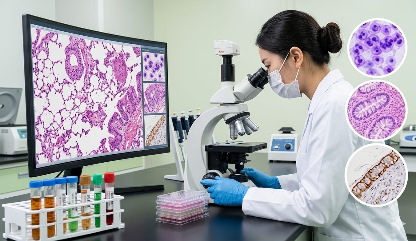
A skin biopsy allows a small sample of tissue to be examined in greater detail. Through histopathological assessment, doctors and pathology professionals can evaluate changes at the cellular and tissue levels.
Skin Histopathology Diagnosis brings together microscopic examination and clinical information to help interpret these changes. Understanding this process can be particularly useful for doctors who encounter complex or unusual skin lesions in clinical practice.
Why Skin Biopsies Matter in Dermatology
A biopsy is not simply a laboratory procedure. It is part of a broader diagnostic process that begins with a clinical question.
Doctors may consider tissue sampling when a lesion has an unusual appearance, persists despite treatment, changes over time, or requires clarification between several possible diagnoses.
Depending on the clinical situation, histopathological examination may contribute to the evaluation of:
Inflammatory skin disorders
Infectious conditions
Autoimmune diseases
Blistering disorders
Benign lesions
Suspicious or neoplastic lesions
The information obtained from the biopsy can add another layer of evidence to the clinical assessment.
Selecting the Right Tissue Sample
The usefulness of a biopsy depends partly on whether the collected tissue adequately represents the area of concern.
The location of the lesion, its clinical characteristics, duration, and suspected diagnosis may influence where and how the specimen is collected.
Relevant clinical information should accompany the specimen whenever possible. This helps the pathology professional understand why the biopsy was taken and what diagnostic questions need to be considered.
Good specimen selection and appropriate clinical information therefore form an important foundation for Skin Histopathology Diagnosis.
What Happens After a Biopsy Is Collected?
Once the tissue reaches the laboratory, it undergoes preparation before microscopic examination.
The specimen is processed so that suitable tissue sections can be produced for examination. Staining techniques can then make different cellular and structural features easier to identify.
At this stage, proper handling is important. A poorly representative or damaged specimen may make interpretation more difficult.
The laboratory process therefore plays an important role in converting a physical tissue sample into information that can be evaluated under the microscope.
What Does the Pathologist Look For?
Microscopic examination provides a much closer view of the tissue than clinical inspection alone.
Depending on the suspected condition, the examination may focus on:
Epidermal architecture
Dermal changes
Inflammatory patterns
Cellular abnormalities
Vascular alterations
Tissue organization
Features associated with infection
Characteristics of benign or malignant lesions
The interpretation usually depends on the overall pattern rather than a single microscopic observation.
This is an important principle of Skin Histopathology Diagnosis, because individual microscopic features may occur in more than one disease.
Understanding Pattern-Based Interpretation
Histopathology involves recognizing relationships between different microscopic findings.
For example, the type of inflammatory cells, their distribution, the skin layers involved, and changes in tissue architecture may all contribute to the interpretation.
A doctor developing an understanding of dermatopathology can therefore benefit from learning patterns rather than trying to associate every individual feature with one specific diagnosis.
This approach encourages more structured thinking when reviewing biopsy findings.
Why Clinical Information Cannot Be Ignored
Microscopic findings should be considered alongside the clinical presentation.
The same histological pattern may have different possible interpretations depending on factors such as the lesion's location, appearance, duration, symptoms, and clinical differential diagnosis.
This is known as clinical-pathological correlation.
Effective communication between the clinician and pathology professional can make the biopsy findings more meaningful. When the clinical question is clearly communicated, the microscopic observations can be interpreted within a more appropriate context.
Skin Conditions That May Require Histopathological Assessment
Dermatopathology has applications across many different categories of skin disease.
Inflammatory Conditions
Inflammatory skin diseases can produce characteristic changes in the epidermis, dermis, or surrounding structures. Histological examination can help describe these patterns and support the broader diagnostic process.
Infectious Diseases
Some infections produce tissue reactions that may provide useful pathological clues. Histopathology can contribute to the assessment, although other laboratory investigations may also be necessary depending on the suspected infection.
Autoimmune and Blistering Disorders
Certain autoimmune diseases affect the structural components of the skin. Detailed pathological examination can contribute to their evaluation, with additional investigations considered when appropriate.
Skin Tumours
Suspicious skin growths may require microscopic assessment to evaluate their cellular characteristics and architecture. Histopathology can provide important information when distinguishing different types of lesions.
When Is Additional Testing Considered?
Routine histological examination is an important part of dermatopathology, but it may not answer every diagnostic question.
Depending on the findings and the clinical concern, additional techniques such as immunohistochemistry or other specialized investigations may be considered.
These methods can provide additional information about specific cellular or tissue characteristics.
Importantly, additional testing is generally selected according to the diagnostic question rather than being automatically required for every biopsy.
Digital Pathology and Dermatopathology Education
Digital pathology has created new opportunities for reviewing and sharing pathology images.
Digitized slides can be examined electronically and can support education, case discussions, review, and collaboration between professionals.
For doctors studying dermatopathology, digital slide resources can also provide opportunities to compare tissue patterns and revisit cases.
Technology can make access to pathological images easier, but the fundamentals of tissue morphology and clinical interpretation remain important.
How Can Doctors Improve Their Understanding of Skin Biopsies?
Doctors who frequently evaluate skin lesions can develop a more structured approach by considering several questions:
What is the main clinical question?
Does the specimen adequately represent the lesion?
What tissue pattern is present?
Which microscopic findings are most significant?
Do the pathological findings fit the clinical presentation?
Is additional testing relevant?
Working through these questions can help connect clinical reasoning with pathological interpretation.
Doctors and medical professionals who want to develop their knowledge in this area can also explore focused educational opportunities such as the Fellowship in Dermatopathology offered by Medical Global Academy.
The broader goal of continued learning is to understand not only what a biopsy shows, but also how those findings relate to the clinical problem.
Frequently Asked Questions
What is Skin Histopathology Diagnosis?
Skin Histopathology Diagnosis involves examining skin tissue microscopically to identify cellular and structural changes and interpreting those findings in relation to clinical information.
Why might a skin biopsy be needed?
A biopsy may be considered when tissue examination can provide additional information about a suspected skin condition or help investigate an uncertain clinical finding.
What information is useful with a skin biopsy?
Details such as the lesion's location, appearance, duration, symptoms, previous treatment, and suspected diagnosis can help provide context for pathological interpretation.
What does a dermatopathologist examine?
A dermatopathologist evaluates skin and related tissue specimens, looking at tissue architecture, cellular characteristics, inflammatory patterns, and other relevant pathological findings.
Why is clinical-pathological correlation important?
Microscopic patterns may sometimes occur in more than one condition. Clinical information helps place those findings in the appropriate diagnostic context.
Does every biopsy require additional testing?
No. Some specimens can be assessed through routine histology, while others may require additional techniques depending on the clinical and pathological findings.
Can digital pathology support learning?
Yes. Digital pathology can provide access to high-resolution tissue images that can be useful for education, case review, and comparison of pathological patterns.
Conclusion
A skin biopsy provides an opportunity to examine changes that cannot always be understood through clinical observation alone. However, interpreting a biopsy involves more than simply looking at a tissue section under a microscope.
Specimen selection, laboratory preparation, microscopic examination, pattern recognition, and clinical-pathological correlation all contribute to the diagnostic process.
Skin Histopathology Diagnosis therefore represents a combination of tissue examination and clinical reasoning.
For doctors interested in dermatology, pathology, or diagnostic medicine, developing an understanding of dermatopathology can make biopsy findings easier to interpret and can support better communication between clinical and pathology teams.
Continued exposure to different cases, tissue patterns, and diagnostic approaches can help medical professionals build a more organized understanding of skin pathology over time.